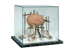
Since 1995 Buyamag Inc Supply Brain Models To Medical Schools
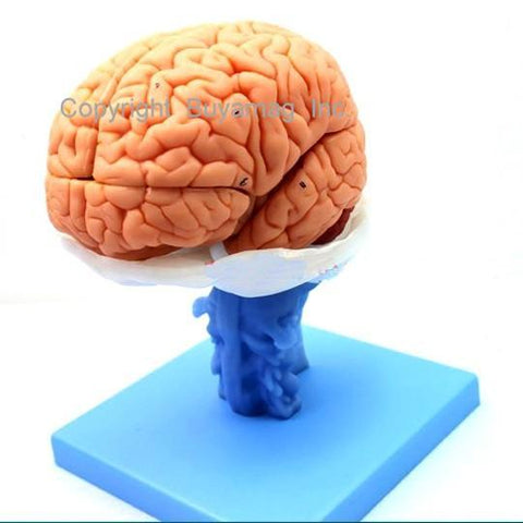
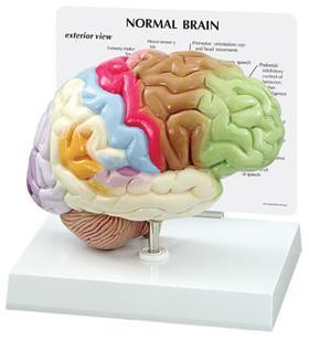
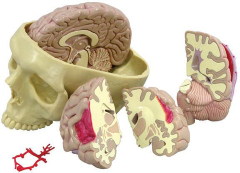
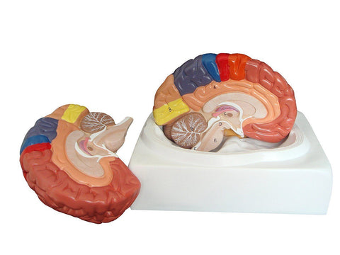
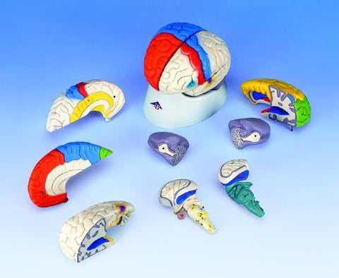
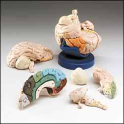
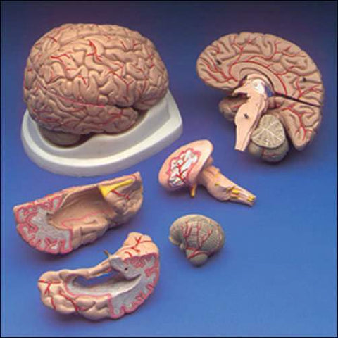
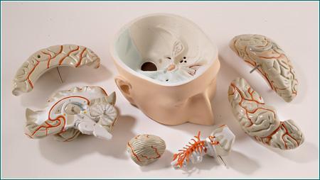
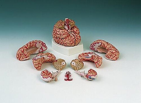
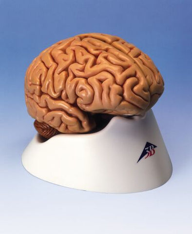
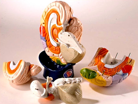
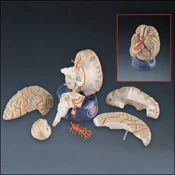
and offer 15 Parts Academy Brain Model Consists of: Cerebral Hemispheres, Corpus Callosum, Insula, Claudate Lenticular Nucleus, Ventricular System, Brain Stem And other 15 components. Quality Brain Related Items

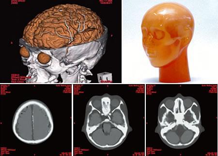
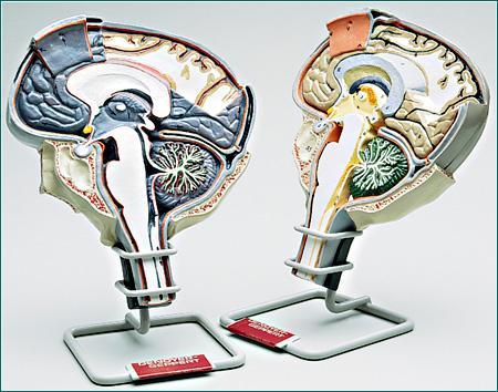
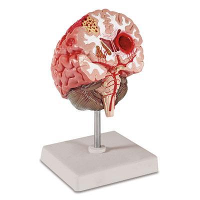
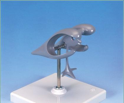
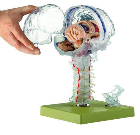
Hands-On experience human brain models showing: Cerebral Aneurysm, Cerebrovascular Hemorrhage And Sagittal Cut Which Exposes The Lateral Ventricle. Help Students Learn How Diagnose And Treat Neurology Disease, Tumor Patient Examination And Treatment.
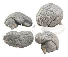
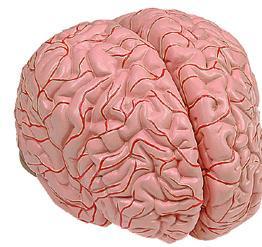
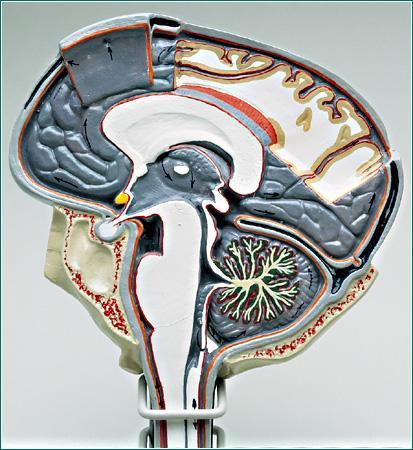
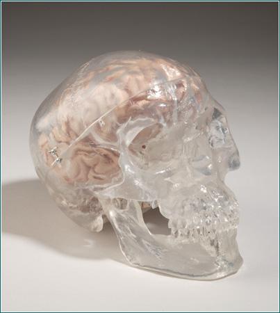
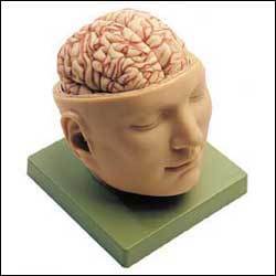
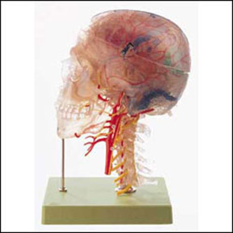
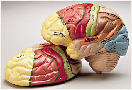
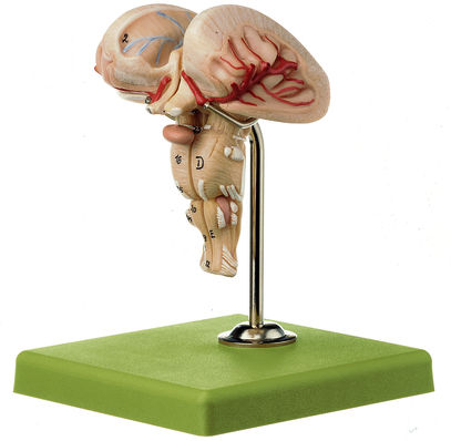
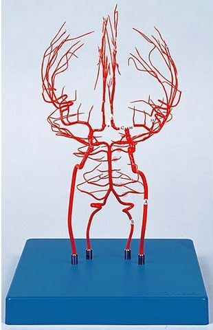
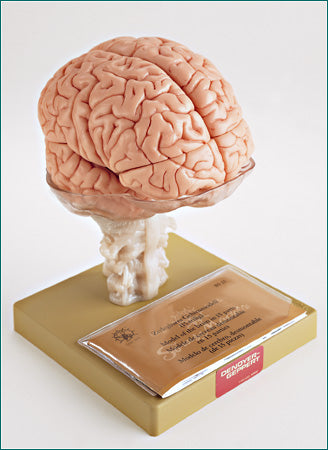
Buyamag Inc established in 1995 in Carlsbad CA 92011 we provide a life-size brain model, divided into hemispheres along the median sagittal plane, this replica depicts the left and right cerebrum, cerebellum, brain stem, medulla and blood vessels. We supply only medical education academy quality anatomical models, used in medical colleges by students and teachers how neurology system working. Also quality brain model neurology science and research. Brain stem medulla and arteries included, pathology sagittal brain vessels circulation with nerves.
Read More »