Your cart is currently empty!
Continue shopping
Cart total : $0.00
www.buyamag.com established in 1995 in Carlsbad CA 92011 we provide Dental Surgical Apex Locator Dentistry Instrument Endo Instrument
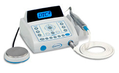
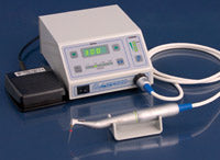
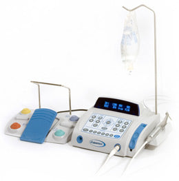
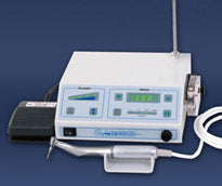
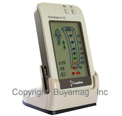
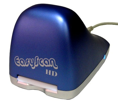
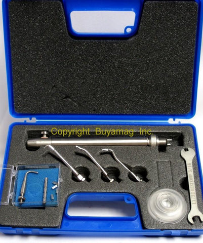
Apex Locator Systems Dental Surgical Rooth Canal Treatment Dental Teeth Implant
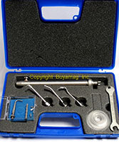
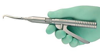
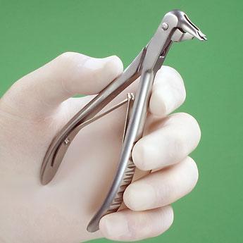
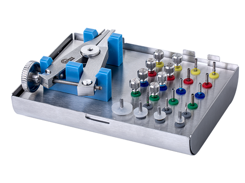
Tooth Endodontic Oral Surgical Easy Cut Gutta Percha Cutting Dentistry Apex Locator Equipment Supply
Dental Easy Cut Surgical Implant Equipment Endodontic Systems Apex Locator Dental Gutta Percha
Read More »
Product successfully added to your shopping cart
Translation missing: en.products.wishlist.added_to_wishlist
Qty:
There are item(s) in your cart