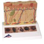
Skin Cancer Model Skin Burn Pathology Model Skin Models Skin Burn Model Skin
Skin Cancer Models Skin Burn Skin Cancer Model Pathology

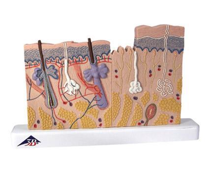
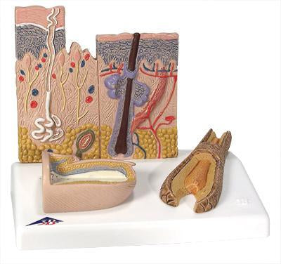
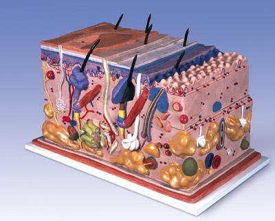
www.buyamag.com established in 1995 in Carlsbad CA 92011 we provide skin model, anatomical models great tools for student education learn study and teaching in medical colleges universities, understand human anatomy, organ function, pathology, and diseases. Obstetric models gynecological simulator are great for female health and child birth study and practicing. Also we provide educational dentistry simulators, Dental Models to dental schools teaching dentistry teeth anatomy, training for gain techniques experience in cosmetic restoration, oral tooth structure, diagnose diseases, pathology treatment procedure. Our dental simulators, manikins phantom head looks and feel patients life-like, give students a chance gain practice experience in dentistry techniques for future professional career. We provide to dental schools orthodontic models, periodontal and implant models so the students can get hands-on oral dentures stomatology training experience, professional knowledge to pass the state board license test, before treat the real patients. We participating in dental courses, curriculums, trade-shows presented by www.buyamag.com
Read More »