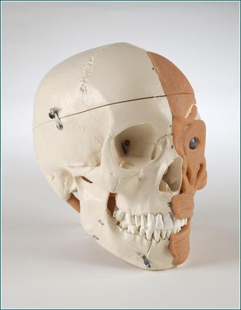
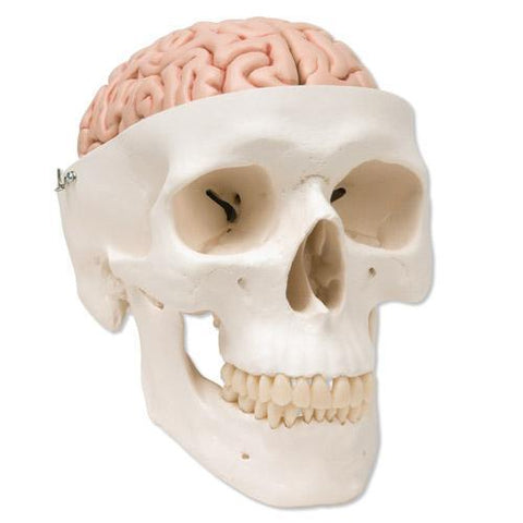
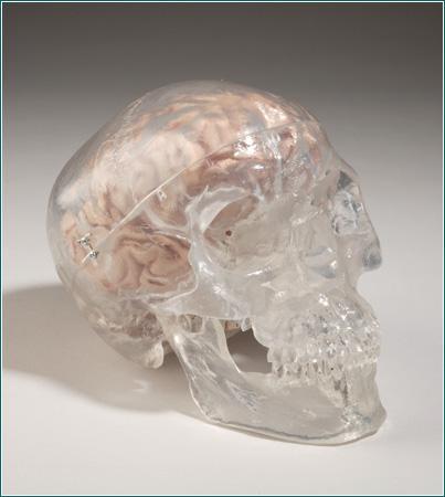
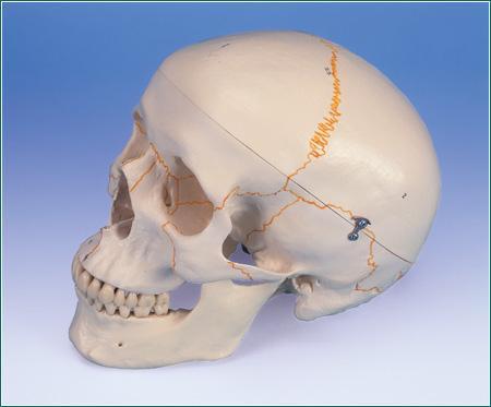
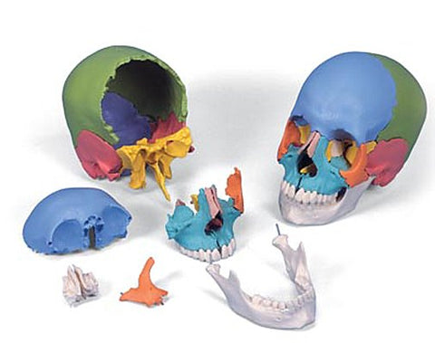
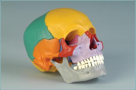
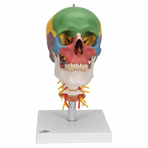
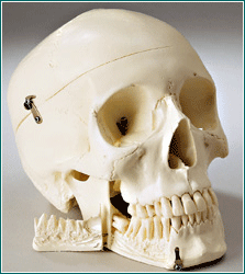
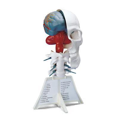
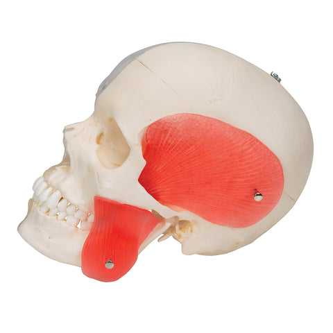
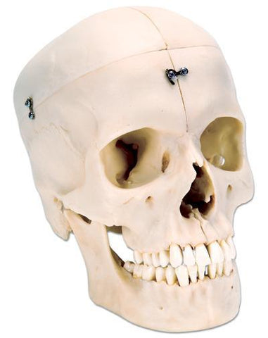
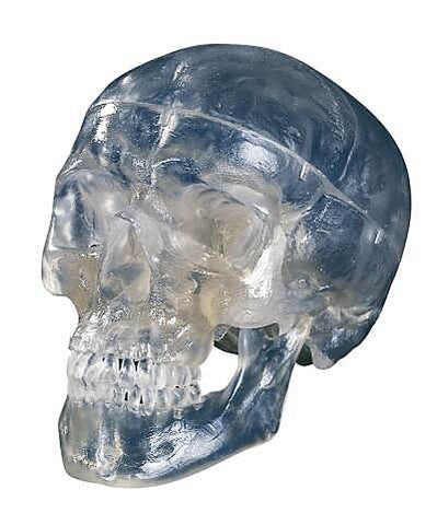
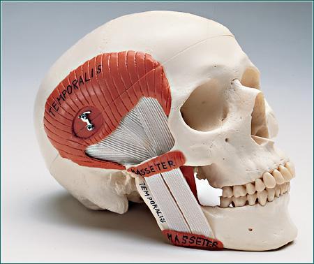
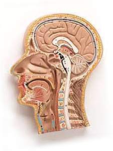
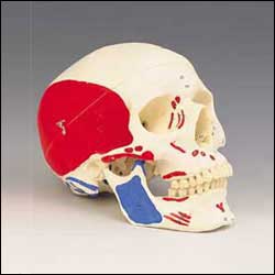
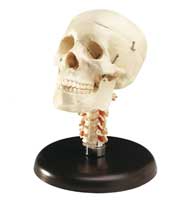
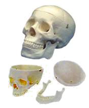
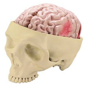
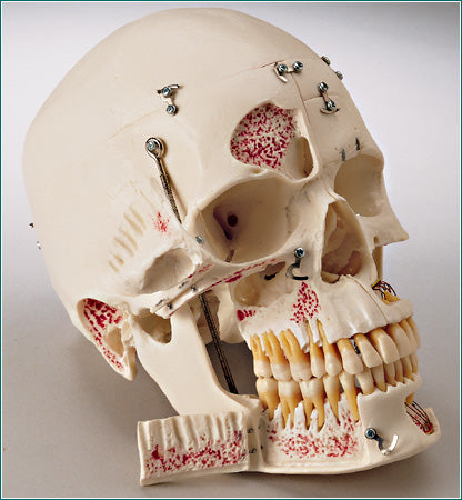
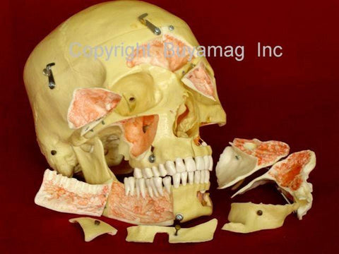
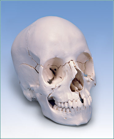
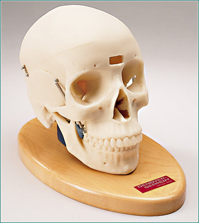
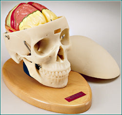
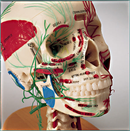
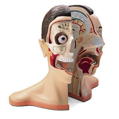
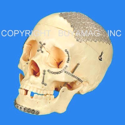
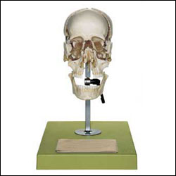
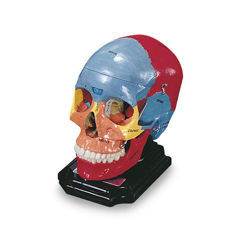
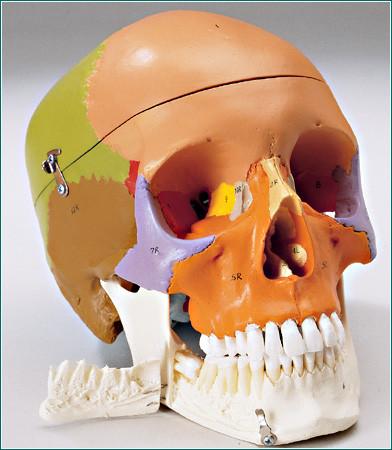
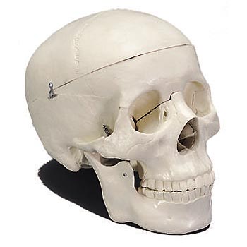
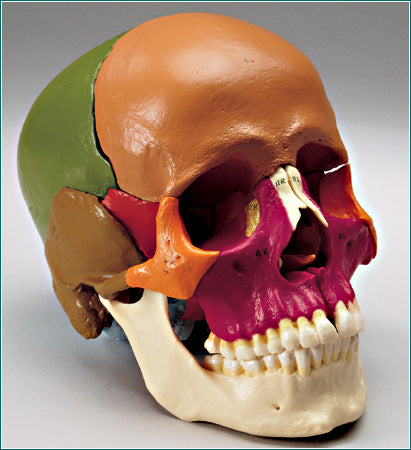
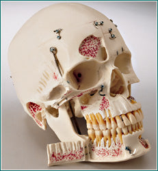
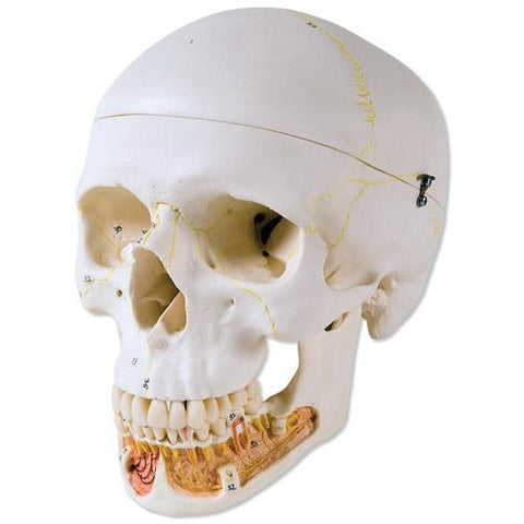
Skull Craniofacial Surgery Cranium Reconstruction Titanium Modular Implants
Devices Simulator Academy Educational Model
Skull
Craniofacial Reconstruction $1499.00
Life-Size Craniofacial Skull
Reconstruction Surgery Simulator. Skull Shows after Surgery,
with Multiple Fractures & Injuries and Titanium Modular Implants
used for Repair. Craniofacial Remodeling Surgery done with Real
Titanium Implants Modular Devices Include: Mash, Plates, Brackets,
Screws.
This Craniofacial Skull Simulator - Model
Teach & Display's Neurosurgery Techniques. Demonstrates a Wide Range
of Titanium Modular Implants Parts & Devices Design and Available in
Skull Craniofacial Reconstruction.
Titanium Implants, Devices & Parts Modular - Multipurpose, and with
Modification can be used in Different Skull Areas. Used in
Neurosurgery Education Programs, Medical Universities,
Neurology Doctors offices for patient presentations and display,
legal presentations.
Craniofacial Scull Professional Academy
Medical Grade
This Educational
Life-Size Adult Human Skull is the Best for Large Auditorium
Educational Purposes. "Exploded" Skull can be Tilted and Locked
in place. 22 Part Disarticulated, Mounted Beauchene Stile;
emphasizes position of Cranial Bones and their relationship to
each other. Cast from the original Beauchene Skull, captures
every details of its bone structure. Design mounted on a
clear plastic base corresponding to the shape of the Skull. Each
of the 16 Bones may be removed for individual study. A
jointed stand attached to a base enables easy maneuvering. Key
card included
Includes:
Frontal, 2 Parietal, 2 Temporal, 2 Zigomatic
2 Nasal, Occipital, 2 maxilla, 2 Lacrimal
2 Nasal Concha, 2 Palatine, Vomer, Ethmoid
Sphenoid Bones, Mandible
Application
Used as a Educational Skull Model, in
Medical Schools and Universities with educational programs,
for students best understand and learn Human Skull Anatomy,
Biology, Function and Pathology. Also used in doctors
offices, laboratories, pharmaceuticals and with legal
presentations
Academy Skull Painted, Cast from
Natural Bone Specimen. Skull with both Styloids, Detailed
suture lines, fissures and fossae, Removable calvarium cap for
neurocranial studies, Open Window into frontal sinuses,
Breakaway maxilla revealing paranasal sinuses, Hinged
mandible highlights perfect dentition, Each bone painted a
different color for easy identification, Names of bones
are hand-lettered. Base Included.
This anatomical painted Skull
with open mandibular is great
quality, articulated, and detailed. Popular Skull in medical and
Dental schools for educational programs and anatomy study, used
in doctors offices and legal presentations.
Strong enough to withstand the
rigors of daily classroom handling. All the major bones are
represented, as well as a full complement of 32 teeth. An
important feature of this skull is that on the lower jaw, the
left canine tooth, the right central incisor, and the 18th
mandibular molar are removable for close study of the roots. The
calvaria is cut and is removable (fastens with clips), so the
interior detail of the cranium can be examined. The lower jaw is
spring held to demonstrate the movement of the jaw. Includes
Human Anatomy Manual.
This anatomical Skull is great
quality, articulated, and detailed. Popular Skull in medical and
Dental schools for educational programs and anatomy study, used
in doctors offices and legal presentations.
Academy
Classic Skull Model 14 Take A - Part Elements
$899.00
Skull with 14 take-apart bone elements. An
invaluable exercise in mastering the orientation and complex
relationships of the bones comprising the adult human skull.
Consisting of 14 separate elements, the
skull can be quickly Dismantled and then Reassembled by means of
flexible snap-together plugs. Sutures have been simplified to
facilitate assembly and disassembly. This version of the
Take-Apart Skull is further enhanced through the use of hand
painted contrasting colors to distinguish the individual bones
which together form the skull
This Life-Size Two the Same Skulls has one
difference - Color Painted or Not Color
Painted. But they share same features: snap plugs
and let your students quickly separate our modular skull
into its 22 individual elements. This unique Human Skull Model
is highly detailed, articulated, dessectable of 22 individual
bones that gradually grow together as the development proceeds.
The new design Skull Kit Model is a natural cast and makes the
complex structure of the skull, bones connection easy to
understand, since it can be disassembled into its 22 individual
bones!. The individual bones can be reassembled by means of
inconspicuous, stable connectors attached at the slightly
simplified skull sutures. The 22 bones are depicted in 9
different didactic colors, detail hand painted so that the
individual skull bones are easy to distinguish. Each pair of
bone plates has the same color. This Skull is Irreplaceable tool
in study Human Skull anatomy and development. Used in medical
schools educational programs by teachers and students, helps
easy understand human anatomy and pathology. Used in doctors
offices, research labs, medical schools, legal presentations.
Two Version 22 parts Skulls available: Color Bones and No color
Bones.
Human Skull 31 piece 3D. Stand and instructions
included. Mounted on a stand. Used in Medical Educational programs
for students understand Human Anatomy and pathology. Also doctors
offices for patience presentations and for legal presentations.
Weight: 1 Lb.
Dental Skull Model With Display Case Deluxe Academy
Dental Skull Academy & Demonstration Display
Case $1599.00
This replica of the Human Dental Skull Deluxe
Academy is one of
exceptional quality and contains amazing Anatomical Educational Detail. This Unique Dental
Education Demonstration Skull has 6
Opening Folders-Flaps, and 10 Parts
Dessectable
to reveal inside the Skull.
Cast from a specially prepared Natural Specimen, this
incomparable Teaching Tool incorporates the following features:
the Calvaria detaches, the remainder of the skull divides into
halves along the Mid-Sagittal plane. The Perpendicular plate of
the Ethmoid and Vomer is hinged to expose the Sphenoid Sinus.
This replica of the human Skull is of an exceptional quality.
The Skull Cap is removable and the base of Skull is
Mid-Sagitally divided. The Frontal Sinus, Perpendicular Lamina
and Vomer are fitted with Folders-Flaps which can be opened to
view the Lateral Nose wall and Sphenoidal Sinus.
On the Left Half : Temporal Bone
detaches • Maxilla is sectioned open revealing the Alveolar
Blood Vessels and Nerves • Frontal Bone has a hinged flap into
the Frontal Sinus. On the Right Half:
the Temporal Bone is opened to reveal the
Sigmoid Sinus, Facial Nerve Canal and the Semicircular
Ducts. Additional opening Folders are located at the
Maxillary Sinus and the right half of the Mandible, so that the
Dental Roots of the Premolars and Molars of the Lower Jaw can
also be viewed. All 32 teeth are individually removable.
Color is used to highlight the sinuses and Semicular Canals, and
to Trace Cranial Nerve Tracts and the Arteries and Meningeal
Sinuses within the Cranium.
This outstanding Demonstration Dental Scull Model 10 part and 6 Opening
Folders design to demonstrate inside details, with natural
occlusion and the individual removal, and replacement of each
tooth also make this Skull especially unique and interesting for
Dentists, Doctors of Neurology, any Medical Schools, and
students educational programs. Additional flaps are located
at the maxillary sinus and the right half of the mandible, so that
the dental roots of the premolars and molars of the lower jaw can
also be viewed. The natural occlusion and the individual removal and
replacement of each tooth also make this skull especially
interesting for dentists. The Anatomy of the human skull is uniquely
displayed in this replica. Included:
Clear Plexiglas
Cover ( hard to brake) on base, for display. This high quality dental skull is a great tool
for students and practicing dentists alike. Or for Doctor - Patient
Demonstration Display Tool.
The new BONElike™ Skulls are made
of a new material that allows an absolutely natural reproduction of
even finest anatomical structures for the first time. Bones made of
3B BONElike™ look real, have an absolutely natural feel and almost
exactly the weight of a natural bone.
By combining one transparent and one bony skull half this is the
first model to allow especially teachers of anatomy a topographical
juxtaposition of the structures that cannot be seen in other skull
versions. The right, transparent skull half allows the study of
important anatomical details, such as the location of the paranasal
sinuses. Therefore, in combination with the left, bony skull half, a
direct transfer of the otherwise invisible structures becomes easy
and uncomplicated.
The transparency of the jaw allows an exceptional view onto the
periodontal pockets and roots. The teeth are removable for detailed
studies. In addition, the external masticator muscles (masseter and
temporal muscles) are represented on the bony skull half. To
demonstrate masticator movement, the lower jaw is mounted flexibly.
These features also make the skull especially valuable for dentists.
The skull can be disassembled into both halves of the skullcap and
base of skull, the nasal septum, the complete mandible and both
masticator muscles.
Skull Bisected 6 Parts. Reveals
the hidden complexities of the Sinuses and Nasal Passages. With
the look and feel of real bone, this highly-detailed plastic
reproduction divides mid-sagittally. Especially suited for
examination of the cranial vault, sinuses, nasal passages and
palate, one half includes the nasal septum and complete
mandible. The other half exposes the nasal conchae and nasal
passages.The new BONElike™ Skulls are made of a new material
that allows an absolutely natural reproduction of even finest
anatomical structures for the first time. Bones made of look
real, have an absolutely natural feel and almost exactly the
weight of a natural bone.
This version represents a complete Midsagitally sectioned Skull.
It can be disassembled into both halves of the Skullcap and the
base of Skull, the Nasal Septum and the complete Mandible. To
demonstrate masticator movement, the lower jaw is mounted
flexibly. An excellent skull to study the bony structure and the
complicated anatomy of the human Skull. Here what customers are saying about
BONElike™:
“This model has an “aged” look that makes it appear like a real
specimen. All the teeth are present and fine structures like the
styloid process, sphenoidal spine and pterygoid hamulus are all
pristine. These fragile structures usually break shortly after
students get their hands on a skull. However, in their plastic
state they are stronger and likely to withstand the rigors of
use better than the original. This is also true of thin areas
such as the lamina papyracea and other parts of the orbital
wall, which, though recognizable on the model, are unlikely to
break as readily as a natural specimen would. One could say that
the model has gained strength and durability at, perhaps, some
small trade-off with realism, but this is exchange is well worth
making for primarily a teaching tool.”
-David Rapaport, Ph.D., Division of Anatomy, Department of
Surgery, University of California, San Diego
Size: 17" x 17" x 11"
This 3-part life-size can make an Forensic
Science Tool, Attractive
Medical Present or Medical Academy Educational Tool. High-quality original cast of real
Human Skull.
Use this "Antique" Skull to study Interior Exterior structures that otherwise
are visible only through x-ray images. It is used by medical schools
educational programs and students, doctor's offices, patients and
legal presentations.
Features
Handmade from hard, unbreakable plastic
Highly accurate representation of the fissures,
foramina, processes, sutures etc.
On 2 pring-mounted Lower Jaw.
2 Lower Teeth are not removable.
Disassemble into at least 2 parts for detailed
studies
Functional Skull Musticator muscles TMJ Demonstration 2 Part
$399.00
Skull 2 part. The Masticatory Muscles (masseter,
temporal, medial and lateral pterygoid muscles) are represented as
elastic bands. Medical Quality Skull has been enhanced to
graphically demonstrate the functioning of the temporman-dibular
joint and the associated TMJ Syndrome.This model is suited to
demonstrate the function of the Masticator Muscles with jaw
occlusion, the initial stage of jaw opening and the movements of the
mandible to the side and front. The Skullcap is removable, Perfect
for patient education, allied health training and medical-legal
documentation, as well as anatomy studies. Functional
synthetic jaw muscles, consisting of resilient colored plastic and
elastic bands, reproduce the Temporalis and masseter muscles on the
right side, the lateral and medial Pterygoids on the left.
Hand-lettered labeling identifies each muscle. Highly accurate
representation of the fissures, foramina, processes, sutures etc
An excellent detailed, hand painted, heavy-duty fiberglass
cross-section of the head and neck. Bones, brain, larynx, nose,
gullet and buccal cavity are numbered. A corresponding key card
is included. Popular in medical schools, students educational
programs for easy understand anatomy, pathology and diseases and
physiology of head area. Used in doctors offices for patience
presentations. Used for legal presentations.
DISCLOSURE
Always consult with your physician before using these Devices & Products. The Buyamag
Inc. does not make any diagnosis or give medical recommendations. These products are not intended to cure, heal, or prevent diseases.
WE do not take back, exchange or refund for Electronic Devices included:
EMS, Tens Machines.
Do Not
Use TENS, EMS (or other devices, machines ), Magnets if you are
fitted with Pace Maker, DEFIBRILLATOR or During Pregnancy, Suspected or
Diagnosed Heart Problems & Epilepsy or other diseases. The medical evaluation of your physician is advised at
all times. Any self-help application is the responsibility of the
user. Magnets can erase video, music, and computer data. Buyamag
Inc.
does not make or imply any medical claims on products described.