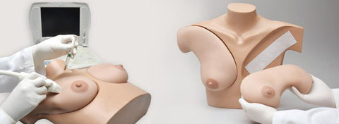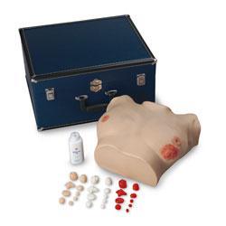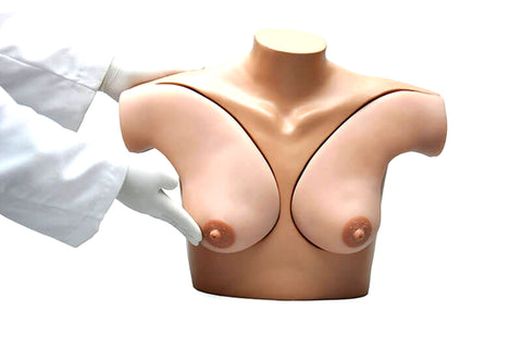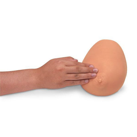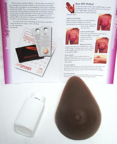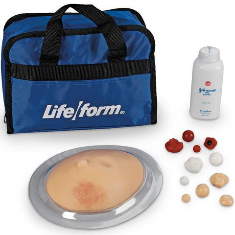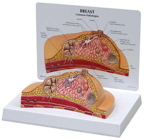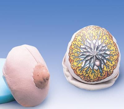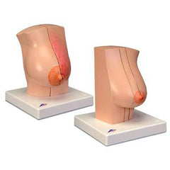
Female 2 Breasts anatomical models, and Chest wall: milk-giving Right breast with representation of an inflammation (mastitis) and non-milk-giving Left breast with representation of various diseases. The Model consists of two Breasts: a milk-giving Female right breast with the surrounding chest wall area, and a non-milk-giving female left breast with the surrounding chest wall area. Both parts of the model have a sagittal cut. The cut surfaces show the tissue of the breast gland as well as the deeper-lying anatomical structures such as the muscles, ribs, costal pleura, pulmonary pleura and lungs. A breast gland inflammation (mastitis) is represented on the right breast and various other diseases on the left breast. The Right Breast: and chest wall are separated into two halves by a sagittal cut in the mammary line and these are held together by magnets. The cut area of the right half shows healthy breast tissue while the cut area of the left half shows the changes to the breast gland when inflamed (mastitis). The exterior of this half shows the associated pathological changes that are visible from the exterior. The Left Breast: and chest wall are divided into three parts by a sagittal cut in the mammary line and an additional internal sagittal cut next to the areola and these are held together by magnets. At the external part of the breast, the skin of the breast is windowed to view the regional lymph nodes. The cut surface of this half show the healthy anatomical structures. The cut surfaces of the middle breast cut show the various diseases of the breast gland. A benign tumour (fibroadenoma) and 2 cysts are represented on the external cut surface. The inner cut surface shows a pathological proliferation of the breast connective tissue (mastopathy). Two malignant tumours are represented in the breast gland on the cut surface of the inner part of the breast. One of these has extended into the chest wall.
Breasts Two Models Features:
- Right and Left Breasts & Various Diseases
- Right Breast: milk-giving breast, 2 sagittal Cuts in Mammary Line
- one half - healthy breast, other half shows mastitis, pathological changes
- Left Breast: non-milk-giving Breast & various Diseases, 3 sagittal custs mammary line, internal sagittal cut next to the Areola, Limph Nodes, healthy
- anatomical structures.
- Middle cut shows various diseases: Benign tumor (fibroadenoma), 2 systs, pathological proliferation (mastopathy), Two malignant tumours