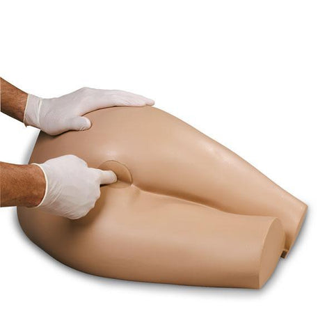

Pre-operative teaching with this Ostomy Simulator allows patients and their families to begin learning about Ostomy prior to surgery, at a time when they are less distracted—reducing anxiety.
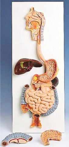
Seeing the 3D digestive and urinary tracts and visualizing the location and function of the various organs is essential to learning, especially in those cases where cognitive processes or language may be an obstacle.
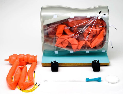
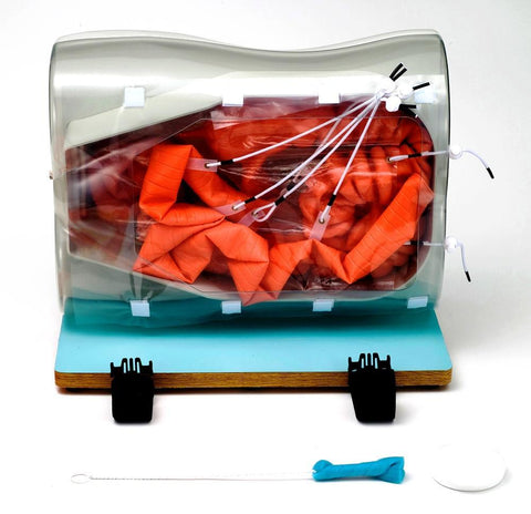
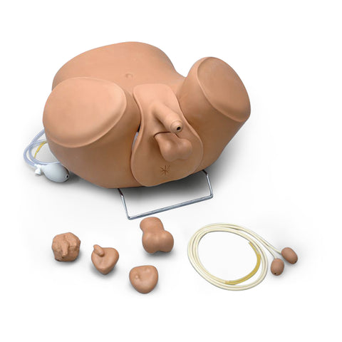
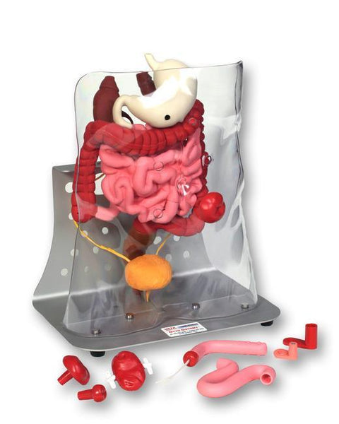
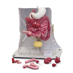
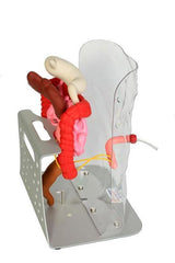
Modeled from an actual patient’s CT scan, Otto Ostomy™ will help your patients and their families become more knowledgeable about what to expect, while demonstrating how ileostomies, urostomies and colostomies function. Otto Ostomy™ has a clear torso shell with four openings for the insertion of stomas.
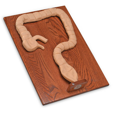
The torso shell is easily removed for teaching or to facilitate in accessing and manipulating the intestines. The color coded organs displayed include: Stomach, Small intestine, Large Intestine, Rectum, Kidneys, Ureters, Bladder. The large intestine, small intestine, rectum and bladder are all removable to aid in teaching procedures where these organs will be removed. The ureters can be removed from the bladder and reinserted into the ileal conduit to show how a urostomy functions.
The flexible small and large intestines can be separated and attached to the backside of the stoma in the torso shell to demonstrate either an end or loop stoma. The large intestine can be separated at four different locations: Ascending colon, Transverse colon, Descending colon, Sigmoid colon. The Torso Shell is easily removed for teaching and to facilitate in accessing and manipulating the Stoma, Organs, Intestines. The flexible Small and large Intestines can be separated in 7 locations and attached to the back side of a Stoma placed in the clear Torso shell to demonstrate various simulation scenarios.
- Enables proper teaching and understanding of Ostomy Procedure
- Increases nurses and patients’ understanding of conditions and adjustments which may be necessary to achieve a satisfactory standard of life with new stoma
- Clear ( see through ) Torso shell with four openings for insertion of stomas
- Shell is easily removed for teaching or to access/manipulate intestines
- Organs are color-coded for easy identification
- Large and small intestines, rectum and bladder are removable to aid in teaching procedures where these organs will be removed
- End or loop stomas can be demonstrated
- Large intestine can be separated at four locations: Ascending colon, Transverse colon, Descending colon, and Sigmoid colon
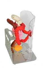
Accessories Include:
- Anodized aluminum base, clear torso shell & 4 openings for stoma placement
- Stomach, small intestine, large intestine, rectum, kidneys, ureters & bladder
- Ileal conduit, two sections of small intestine
- Two adaptor connectors (to demonstrate loop stoma with either the small or large intestine)
- Four stomas: 7/8” diameter, 2” diameter, 7/8” with two 3” stents (to be used with the ileal conduit)
- Loop stoma with rod, & user manual.
- Size ....... 17" x 10" x 16", Wt .......... 9Lbs
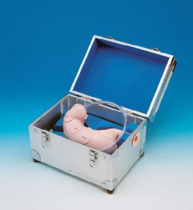
- Carrying Case --------------- Optional.