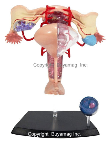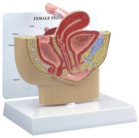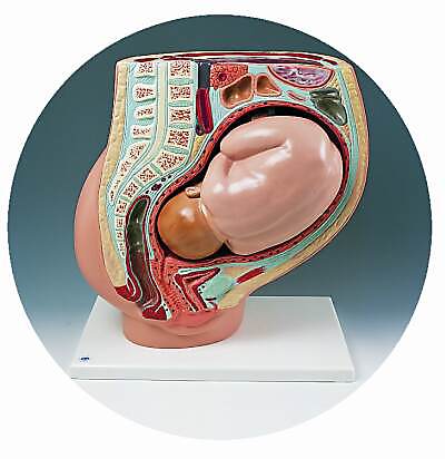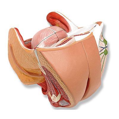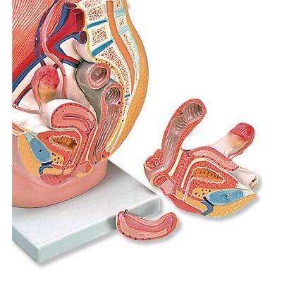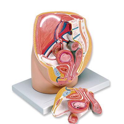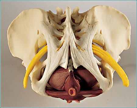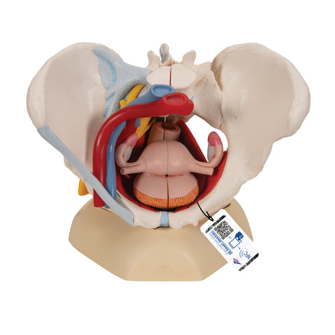
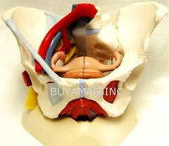

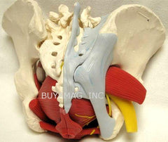
This Six Part of a Female Pelvis Life-Size Academy Model represents detailed information about the Topography of Bones, Ligaments, Vessels, Nerves, Pelvic Floor Muscles and Female Pelvic Organs. It presents the Whole Pelvic Floor with partially Removable Midsagitally Sectioned External Anal Sphincter, External Urethral Sphincter, deep and superficial Transverse Perineal and Bulbospongiosus. Rectum, Uterus with Fallopian Tubes and Ovaries and Vagina are also removable and can be disassembled into both halves by Midsagital Section. The right Pelvic Half demonstrates the divisions and topographical anatomy of the common iliac artery, the external and internal artery and also of the common iliac vein and the external iliac vein. The right sacral plexus, right sciatic nerve and right pudendal nerve are also shown. Bones and ligaments presented: Two hip bones, the pubic symphysis, the sacrum and the coccyx, the fifth lumbar vertebra with intervertebral disc. A midsagital section through the fifth lumbar vertebra, sacrum and coccyx, allow both halves of the pelvis to be disassembled revealing a part of the cauda equina in the vertebral canal. The Left half of the fifth lumbar vertebral body is removable. The right half of the model shows following pelvic ligaments: inguinal ligament, sacrotuberous ligament, sacrospinous ligament, anterior sacroiliac ligaments, iliolumbar ligament, anterior longitudinal ligament, interosseous sacroiliac ligament, posterior sacroiliac ligament and obturator. Both Left and Right Sides holds together with 8 powerful Neodymium Magnets to insure quick & easy assembly of the Pelvis. The organs are a softer, more rubbery plastic that is pliable material. Plastic Presentation Stand included. Size: 8 x 11 x 8
Female Pelvis Features:- Topography of Bones Ligaments Vessels Nerves Pelvic Floor Muscles & Female Pelvic Organs
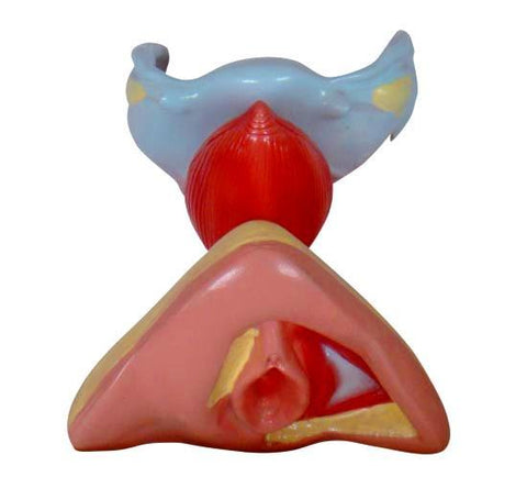
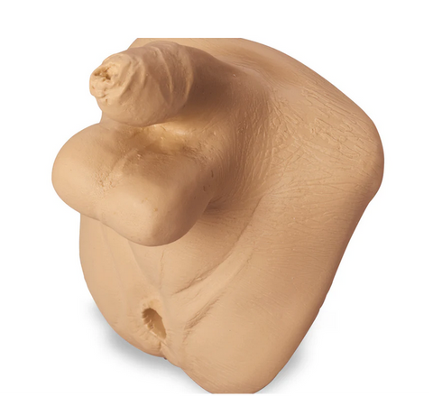
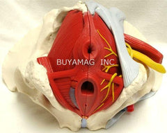
- Whole Pelvic Floor with Partially Removable Midsagitally Sectioned External Anal Sphincter, External Urethral Sphincter,
- Deep and superficial Transverse Perineal and Bulbospongiosus.
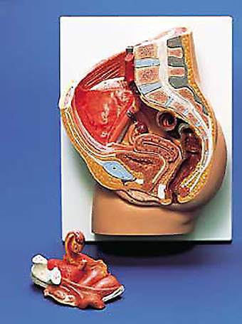
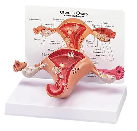
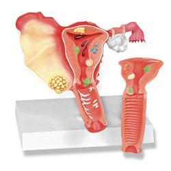
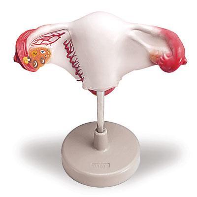
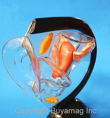
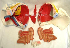
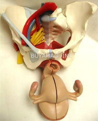
- Rectum, Uterus with Fallopian Tubes and Ovaries and Vagina are also removable and can be disassembled into both halves by Midsagital Section.
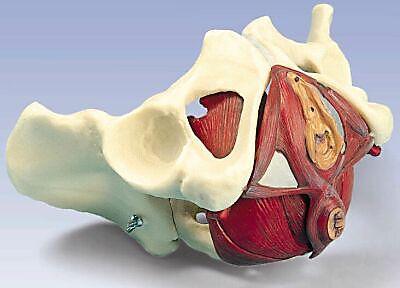
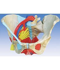
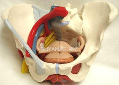
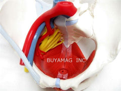
- Right Pelvic Half demonstrates: the divisions and topographical anatomy of the common iliac artery, the external and internal artery and also of the common iliac vein and the external iliac vein
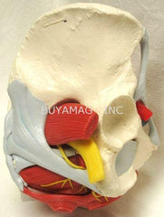
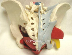
- Sacral plexus, right sciatic nerve and pudendal nerve, Bones and ligaments presented: Two hip bones, the pubic symphysis, the sacrum and the coccyx, the fifth lumbar vertebra with intervertebral disc. A midsagital section through the fifth lumbar vertebra, sacrum and coccyx, allow both halves of the pelvis to be disassembled revealing a part of the cauda equina in the vertebral canal.
- The Left Pelvic Half demonstrates: the fifth lumbar vertebral body is removable. The right half of the model shows following pelvic ligaments: inguinal ligament, sacrotuberous ligament, sacrospinous ligament, anterior sacroiliac ligaments, iliolumbar ligament, anterior longitudinal ligament, interosseous sacroiliac ligament, posterior sacroiliac ligament and obturator.
- Both Left and Right Sides holds together with 8 powerful Neodymium Magnets to insure quick & easy assembly of the Pelvis. The organs material are a softer, more rubbery plastic that is pliable material
3 years warranty
Anatomical Models