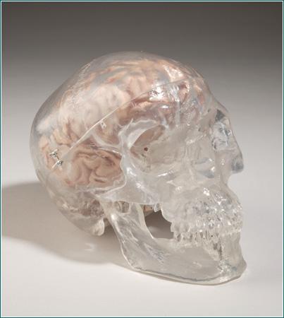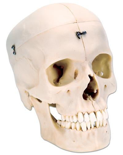

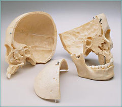
Skull Model Bisected Reveals Hidden Sinuses Nasal Passages
Buyamag INC
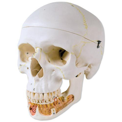
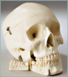
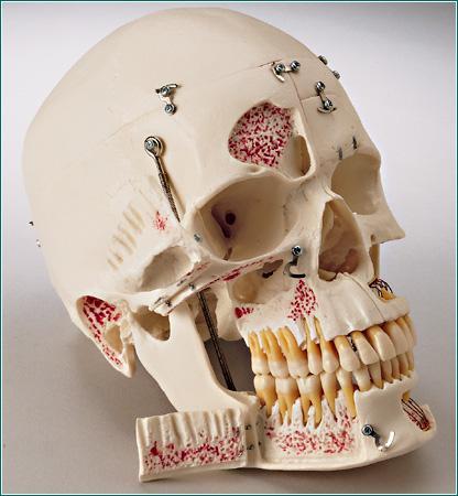
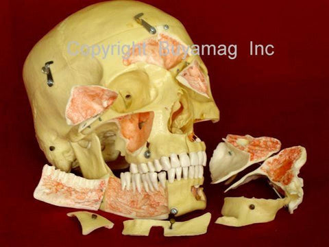
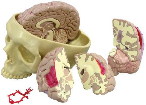
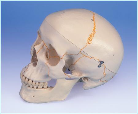
Skull model bisected 6 Parts. Reveals the hidden complexities of the Sinuses and Nasal Passages. With the look and feel of real bone, this highly-detailed plastic reproduction divides mid-sagittally. Especially suited for examination of the cranial vault, sinuses, nasal passages and palate, one half includes the nasal septum and complete mandible. The other half exposes the nasal conchae and nasal passages.The new BONElike™ Skulls are made of a new material that allows an absolutely natural reproduction of even finest anatomical structures for the first time. Bones made of look real, have an absolutely natural feel and almost exactly the weight of a natural bone.
This version represents a complete Midsagitally sectioned Skull. It can be disassembled into both halves of the Skullcap and the base of Skull, the Nasal Septum and the complete Mandible. To demonstrate masticator movement, the lower jaw is mounted flexibly. An excellent skull to study the bony structure and the complicated anatomy of the human Skull.
Here what customers are saying about BONElike™:
“This model has an “aged” look that makes it appear like a real specimen. All the teeth are present and fine structures like the styloid process, sphenoidal spine and pterygoid hamulus are all pristine. These fragile structures usually break shortly after students get their hands on a skull. However, in their plastic state they are stronger and likely to withstand the rigors of use better than the original. This is also true of thin areas such as the lamina papyracea and other parts of the orbital wall, which, though recognizable on the model, are unlikely to break as readily as a natural specimen would. One could say that the model has gained strength and durability at, perhaps, some small trade-off with realism, but this is exchange is well worth making for primarily a teaching tool.”
-David Rapaport, Ph.D., Division of Anatomy, Department of Surgery, University of California, San Diego