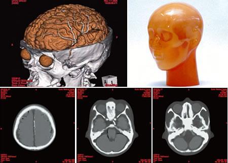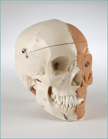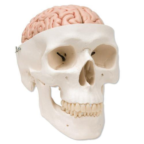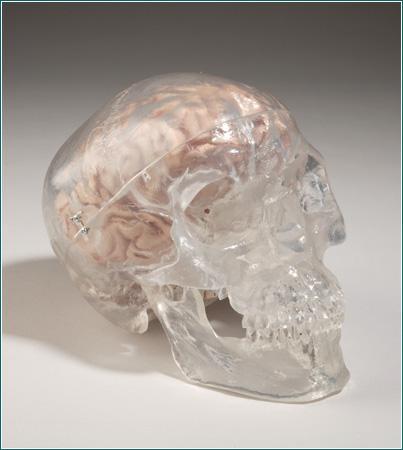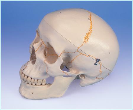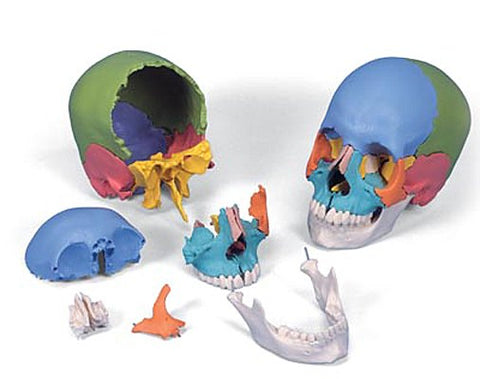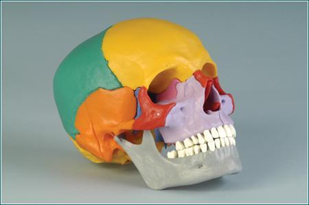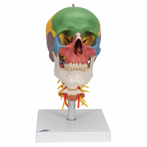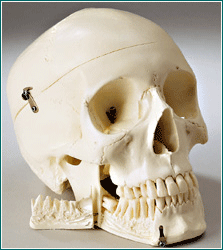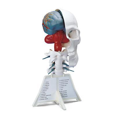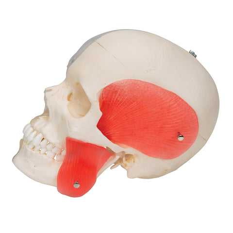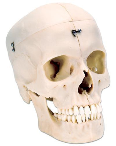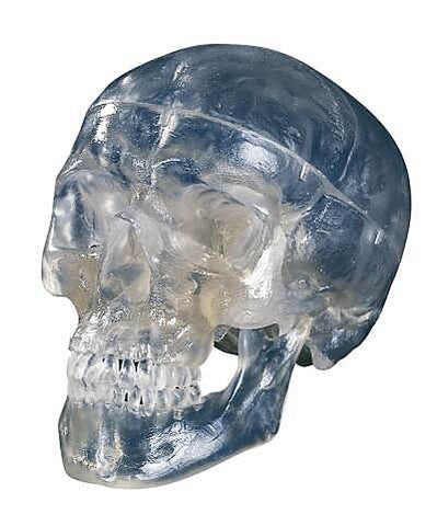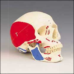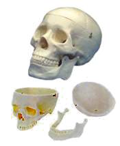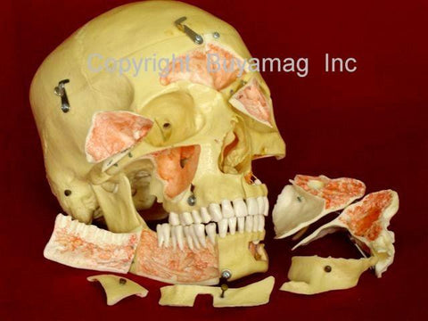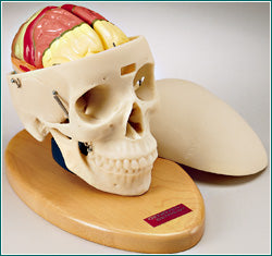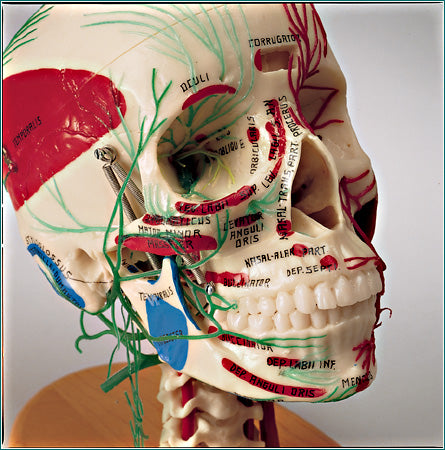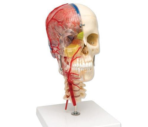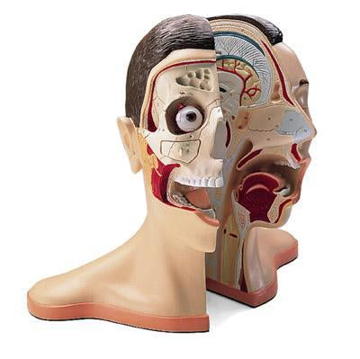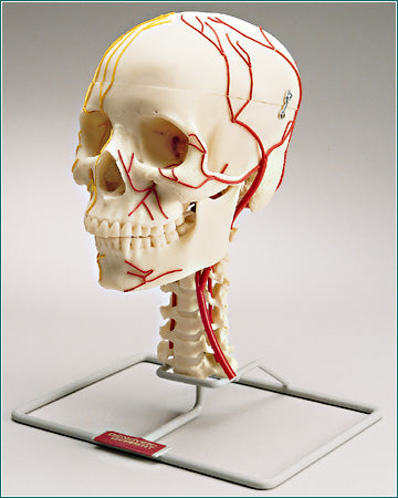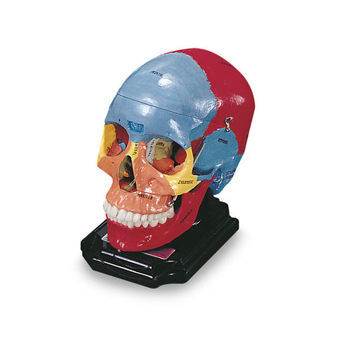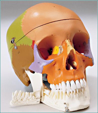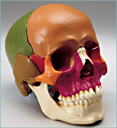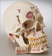
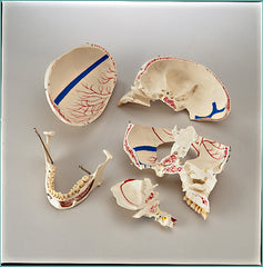
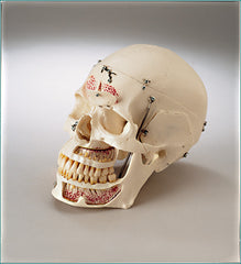
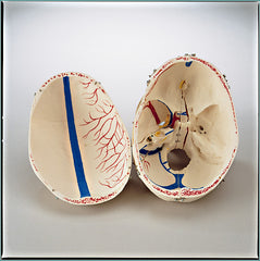
This unique dental skull model dental demonstration has 6 Opening Folders-Flaps, and 10 Parts Dessectable to reveal inside the Skull. Cast from a specially prepared Natural Specimen, this incomparable Teaching Tool incorporates the following features: the Calvaria detaches, the remainder of the skull divides into halves along the Mid-Sagittal plane. The Perpendicular plate of the Ethmoid and Vomer is hinged to expose the Sphenoid Sinus. This replica of the human Skull is of an exceptional quality. The Skull Cap is removable and the base of Skull is Mid-Sagitally divided. The Frontal Sinus, Perpendicular Lamina and Vomer are fitted with Folders-Flaps which can be opened to view the Lateral Nose wall and Sphenoidal Sinus. On the Left Half : Temporal Bone detaches • Maxilla is sectioned open revealing the Alveolar Blood Vessels and Nerves • Frontal Bone has a hinged flap into the Frontal Sinus. On the Right Side: the Temporal Bone is opened to reveal the Sigmoid Sinus, Facial Nerve Canal and the Semicircular Ducts. Additional opening Folders are located at the Maxillary Sinus and the right half of the Mandible, so that the Dental Roots of the Premolars and Molars of the Lower Jaw can also be viewed. All 32 teeth are individually removable. Color is used to highlight the sinuses and Semicular Canals, and to Trace Cranial Nerve Tracts and the Arteries and Meningeal Sinuses within the Cranium. This outstanding Demonstration Scull Model 10 part and 6 Opening Folders design to demonstrate inside details, with natural occlusion and the individual removal, and replacement of each tooth also make this Skull especially unique and interesting for Dentists, Doctors of Neurology, any Medical Schools, and students educational programs.
Size: 12" x 9.5" x 7.7"
Weight: 1.1kg
LIFETIME WARRANTY!